


External Fixator Systems: A Definitive Guide for Orthopedic Surgeons
from web site
In the hierarchy of fracture fixation strategies, the external fixator occupies a unique and irreplaceable position. Unlike internal fixation devices that are implanted within the body, external fixators achieve fracture stability through pins or wires inserted percutaneously into bone fragments and connected to an external frame that spans the fracture — providing mechanical control from outside the skin. This approach enables fracture stabilisation in situations where internal fixation would be dangerous, impractical, or premature.
Core Principles of External Fixation
External fixation achieves fracture stability by the same fundamental biomechanical principle as internal fixation — controlling the relative movement of fracture fragments — but through a completely different mechanical pathway. Pins or wires trans-fix the bone fragments, passing through the skin surface into the bone at specific locations calculated to provide adequate purchase while avoiding critical neurovascular structures. These pins are then connected to an external frame — typically constructed from aluminium alloy or carbon fibre rods and clamps — that holds them in the correct spatial relationship, preventing displacement of the fracture fragments during patient movement and load application. The mechanical stiffness of the external fixator construct, which determines how much interfragmentary movement occurs under physiological loading, is adjustable through the frame configuration, pin number, and pin diameter.
Indications: When External Fixation Is the Right Choice
External fixation is indicated across several distinct clinical scenarios where its unique properties provide advantages over alternative fixation methods. Damage control orthopaedics in polytrauma patients uses temporary external fixation to rapidly stabilise long bone and pelvic fractures in patients too physiologically compromised for definitive internal fixation, allowing the surgical team to address life-threatening injuries before returning for definitive skeletal reconstruction. Open fractures with extensive soft tissue contamination benefit from external fixation because it avoids placing hardware in a contaminated wound. Fractures with severe comminution that precludes internal fixation and fractures in infected bone where internal hardware would risk infection propagation are also appropriate external fixation indications.
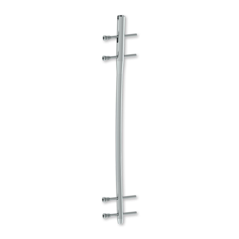
Femoral Interlocking Intramedullary Nail — Orthopedic Fixation Products — Youbetter Medical (youbettermed.com)
Frame Configurations: Uniplanar, Biplanar, and Circular
External fixator systems are available in three primary configurations, each offering different levels of stability and versatility. Uniplanar fixators use pins arranged in a single plane and connected to a linear bar, providing stability primarily in the plane of the frame — appropriate for simple fractures in cooperative patients. Biplanar fixators add a second plane of fixation, dramatically increasing construct stability for more complex fracture patterns. Circular fixators — of which the Ilizarov ring fixator is the archetype — use fine wires tensioned under high load through circular rings connected by threaded rods, enabling precise three-dimensional control of fragment position that makes them powerful tools for complex deformity correction and limb lengthening in addition to fracture management.
Pin Care and External Fixation Complication Management
The interface between the fixation pin and the skin surface is the primary site of external fixation complications. Pin tract infection — ranging from superficial cellulitis around the pin exit wound to deep osteomyelitis at the pin-bone interface — is the most common complication of external fixation, occurring in 5 to 30% of cases depending on the fracture type, pin care protocol, and duration of external fixation. Meticulous pin care, including daily cleaning with approved antiseptic solution and monitoring for early signs of infection, reduces the frequency and severity of these complications substantially. The quality and surface finish of the pins themselves also influences infection rates — polished pin surfaces with accurate thread form reduce micro-motion at the skin interface that promotes bacterial ingress.
