20 Reasons You Need To Stop Stressing About Health Check Up
from web site
Before doing any kind of radiological treatment, the factors that the radiologists must take of are: Defense from the radiation, The impacts of the radiations on the human body, Carrying out Properly, Correct Interpretation of the examination.
There is range of Radiology procedures as well as every examination is executed in different ways.
CT scan- a diet regimen is to be complied with purchased. The client is provided with a contrast agent by mouth, intravenously or rectally. It is a painless test. In this the patient is stocked a movable table and is moved inside a rounded device and also asked to hold the breath for a while. The entire process takes a time of about 15 to half an hour.
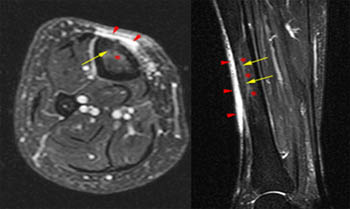
MRI check- it is additionally a painless evaluation in which is positioned under a big round magnet that has a very high electromagnetic field. It is a risk-free process Radiology treatments. Some preventative measures need to be taken before this test. People with speed maker are not allowed to go extensive this test. The whole process takes about 1 to 2 hrs.
PET check is increased as positron exhaust tomography. It reveals the body metabolic process rather than showing the makeup. Prior to the test the person is provided with radioactive sugar and allowed to take remainder so that the sugar gets distributed completely throughout the body and after that glided inside the scanner to continue the procedure. It takes around 1.5 to 3 hrs to complete.
Angiogram- in this examination a catheter is placed into the artery through which a contrast product is provided. If the tests are to be taken in the early morning after that the client is not enabled to have food as well as drink water after twelve o'clock at night. This examination is a pain-free one and also takes close to about 2 hours.
Ultrasonography- in this acoustic wave with high frequency are usage to imagine inside the body and then gotten by the transducer which is viewed as an image in the monitor. USG is of several kinds. Pelvic stomach, renal etc. In pelvic USG the client is asked to consume 30-to 45 oz of water before the test. This test is additionally pain-free.
Radiology engineers take xrays and also administer nonradioactive products right into patients' blood streams for analysis purposes. Some focus on diagnostic imaging modern technologies, such as computerized tomography (CT) and also magnetic resonance imaging (MRI). Radiologic engineers and technicians, likewise described as radiographers, produce xray films (radiographs) of parts of the human body for usage in diagnosing clinical issues.
They prepare patients for radiology exams by explaining the treatment, removing posts where xrays can not pass as well as positioning patients to make sure that the parts of the body can be properly radiographed.To avoid unneeded radiation exposure, these workers surround the subjected area with radiation defense gadgets, such as lead shields, or restrict the size of the xray light beam with collimation.
Radiology technologists position radiographic tools at the appropriate angle and also height over the appropriate area of a client's body. Making use of tools comparable to a measuring tape, they may gauge the thickness of the section to be radiographed as well as established controls on the xray device to produce radiographs of the suitable thickness, information, and also contrast. They position the x ray movie under the part of the patient's body to be examined as well as make the direct exposure. They then remove the movie and develop it.
After inspecting the movie for high quality, the rad tech will certainly send it to the radiologist for analysis. The individual is launched and told to anticipate the outcomes.
Radiology is another kind of medical specialized which is utilized ti get photos of various parts of the body to discover and also deal with illness. Different imaging methods are made use of by the radiologists as well as one of the most important among them are X-RAY, USG, CT Scan, nuclear medicine, PET and MRI.
There are various kinds of Radiology strategies which are stated as under:-.
X-Rays- it is also called radiographs. There are created by passing x-rays with the person's body which then gets guided to a recording device and more created as an image. One of the most commonly secondhand kind of imaging is the Silver Containing films which is currently changed by Digital radiography. As a result of its availability as well as cheap prices is the most recommended test provided by the physicians.
Fluoroscopy- Angiography or Fluoroscopy are the unique type of an x-ray applications. In this a display and also an intensifier is utilized which help in the development of the picture both this things are connected to a close circuit television. The person as administered with different agents to differentiate between the cells. It is typically made use of to identify lumps or cysts.
Interventional radiology- it is mainly utilized to diagnose and deal with outer vascular diseases, Inferior vena cavern filter positioning, gastrostomy tube positionings, biliary stents and also hepatic interventions in a minimally invasive technique.
Computed Tomography- X-rays is used on top of that with formulas to take picture of the body. It is used for identifying urgent scenarios such as hemorrhage, clots in the arteries of the lungs, appendicitis, and also healing kidney rocks.
Ultrasound-it is made use of to envision the fetus, kidney stone, spleentomegaly and so on it utilized the high frequency sound waves to detect the abnormalities.
Magnetic Resonance Imaging- it makes use of magnetic fields to find the core of the atom within the tissues, then utilizes a radio signals to produce disturbance in the axis of turning of core and also observes the superhigh frequency signal generated And none the less are the nuclear medications imaging which are administered right into the clients consisting of materials which have the affinity for tissues labeled with radioactive tracer.
Preferably, radiology solutions ought to be available 24/7 in medical facilities for the rapid analysis of examinations as well as timely treatment of medical problems. This is particularly essential for emergency situation scenarios where time is essential.
However, this isn't constantly the instance - especially for smaller hospitals, facilities or methods. The arrival of teleradiology has actually made this feasible for these establishments and people, permitting them to give faster, excellent quality client treatment.
Healthcare facility emergency clinic, medical wings, and other extremely critical medical therapy environments constantly need radiological images taken immediately for individuals who experience crashes or major problems that emerge all of a sudden. With teleradiology, doctors are able to enlist specialist radiology services right away to help safeguard a medical diagnosis.
Certain companies are devoted specifically to providing such solutions as well as utilize full time radiologists that are readily available day-and-night. Such solutions can consist of a selection of specialties as well as subspecialties, varying from body imaging, to pediatric radiology, to cardiovascular imaging. Radiologists use the most recent photo archiving and also communications system to get, evaluate and also interpret radiological photos, such as X-rays, MRIs and CT scans.
With the right modern technology and high-speed Web, radiologists can execute these and create both initial and also last reports from their residences, despite the time or day. In some business, records can even be provided in as quickly as 30 minutes. Teleradiology has actually led the way for extra quick diagnosis as well as treatment.
Speed, reliability and also flexibility have actually made this technique gain enhancing popularity among medical centers throughout the nation. Since when it concerns individual treatment, there's no time at all to waste.
A number of methods are utilized to envision body organs either directly or indirectly, Panoramas of medical imaging have broadened explosively recently and also are still establishing quickly.
The most classic as well as well developed method of imaging is radiology, which uses X-rays. Darkness cast on the photosensitive movie by different tissues vary in thickness and this principle is filed a claim against in analyzing the radiographs. Different techniques like ordinary radiography, contrast radiography as well as tomography are employed. Radiological imaging offers details regarding physiological and structural modifications in an body organ, e.g, foreign bodies in the bronchi, combination of the lungs, heart enlargement, abnormalities of bones, and so on. Both the structural irregularities as well as physical functions can be researched by techniques utilizing comparison radiography, e.g, barium swallow, hyperbaric oxygen therapy barium dish follow up, cholecystography, comparison urography, and so on. Angiography elegantly reveals the vascular supply of an body organ. Aside from imagining occlusion and also aneurysms, the vascular pattern gives indirect evidence of tumors, space occupying lesions as well as likewise the functional state of the organ. Angiography has actually been thoroughly used in Cardiovascular, neurological, kidney, hepatic, and other problems. Angiography has actually been made use of with various other approaches like computerized tomography to enhance the resolution of details even more. Angiography has actually been put on the arteries, blood vessels as well as lymphatics.
A brand-new development in the field is interventional radiology in which investigatory or therapeutic treatments are done under radiological control. The method is highly innovative, demanding really great skill and best group work. A couple of timeless examples of interventional radiography are endoscopic backward cholangiopancreatography (ERCP) with elimination of pancreatic or biliary calculi; renal artery expansion through a renal artery catheter and also relief of coronary occlusion using a balloon catheter in the coronary artery.
Radiology is still the standard strategy of imaging because this examination responds to most of the questions. Moreover its universal accessibility and also relatively affordable have aided to make it the most appropriate examination. Though conventional readiography is noninvasive, contrast studies are intrusive in differing levels. The exposure to analysis X-rays, though tiny quantitatively, adds to advancing irradiation gotten by the subject. It is well-known that irradiation of the fetus in utero, specifically throughout very early pregnancy can be harmful to the infant. So also duplicated radiographic research studies can offer cumulative toxicity due to X-rays. Though the dose as well as the area of exposure have actually been significantly minimized in modern machines, this risk should not be ignored and also radiological research studies ought to be taken on just if appropriately indicated. Several radiological strategies are supplemented by the more recent imaging methods like Ultrasonography, isotope scanning, computerized tomography and nuclear magnetic vibration.
