The Microscope
from web site
Most sophisticated microscopes that go up to 1000x come equipped with an Abbe condenser, which can be focused by moving it up and down. The Abbe condenser should be set closest to the slide at 1000x and moved further away as the magnification level gets lower. The left image was dry and the right image was with microscope immersion oil. Notice the difference in image quality and the resolution between the image captured dry versus with immersion oil.
Other scientists did not use his microscopes, as they were difficult to learn to use. Some improvements to the device occurred in the 1730s, but big improvements that led to today's compound microscopes didn't happen until the middle of the 19th century. With these microscopes, though, he made the microbiological discoveries for which he is famous.
Stereo microscope magnification helps sort and visualize peripheral surfaces in three-dimensions which allows for a thorough examination of objects. This means that an object 20mm (2cm, or about 3/4 inch) wide would fill up the whole viewing area at 10x and an object about 6.7mm wide would fill up the whole area at 30x. As you can see, having the highest power may not be best for your particular application.
The ultrasonic linear motors provide a very wide dynamic speed range from microns/second and below, up to more than 100mm/sec. A linear encoder with 100nm resolution is integrated for each axis – this direct position measurement principle eliminates play and backlash. The U-780 XY stage systems come with many software tools, such as PIMikroMove, PI General Command Set , and drivers for LabVIEW. You will need access to a cell phone that enables you at least to zoom manually in and out. You will need a 1-millimeter optical ball lens to magnify the sample.
Because the objective lens is convex, it focuses and directs light into its center. By contrast, the concave shape of the ocular lens serves to spread out the light as it meets the eye, thereby making the image bigger. The condenser is a lens, often implanted into the stage or located just below it, that condenses the light rays from the light source onto the spot that is being examined on the specimen above.
Generally high vapour solvents are used in order to limit contact time with surfaces and cements. Caution is required when dealing with older microscopes if too much solvent is applied then this may affect the integrity of the mountant e.g. cement or balsam in particular. With modern microscopes this is less of a problem due to the use of polyacrylamates and other more resistant substances.
Modern Photonics Microscope Lens Adapters
In the gold-standard case, it is approximately 180 × 180 μm for 1.25 NA, 100 × oil immersion microscope objective. Current WHO standard requires 100 fields of view to provide the final determination of malaria infection. The standard limit of detection is 30 parasites/μl of blood. The system is almost insensitive to the distance between the glass ball and the phone lens.
For starters, a microscope is an optical tool that can be used to view very small and near objects in great detail. Both instruments were invented hundreds of years ago and have been used to make significant advancements in biology, chemistry, and astronomy. The diameter of the lenses of microscopes and telescopes are substantially different. An optical instrument having a magnifying lens or a combination of lenses for inspecting objects too small to be seen distinctly by the unaided eye.
Microscopes use a convex lens to generate extremely magnified images of very small objects. The lens at the end of the simple microscope produces an inverted and magnified image. Biconvex lenses are a simple lens comprising two convex spherical surfaces, generally with the same radius of curvature. … Being a simple lens, biconvex lenses have a broad range of applications including, but not limited to, focussing and control of laser beams, moderate quality imaging and other optical instruments. The VH lens series features high-performance lenses that bring the best observation and imaging out of microscopes.
Cells are building blocks for all living organisms, but they are too small to be seen with our naked eyes. With the aid of microscopes, we can amplify the power to see these tiny cells. There are also many tiny creatures that we can not see with our naked eyes, such as plankton and bacteria. Illustrated in Figure 8 is a schematic drawing of light waves reflecting and/or passing through a lens element coated with two antireflection layers. The incident wave strikes the first layer at an angle, resulting in part of the light being reflected (R) and part being transmitted through the first layer.
Other scientists did not use his microscopes, as they were difficult to learn to use. Some improvements to the device occurred in the 1730s, but big improvements that led to today's compound microscopes didn't happen until the middle of the 19th century. With these microscopes, though, he made the microbiological discoveries for which he is famous.
Stereo microscope magnification helps sort and visualize peripheral surfaces in three-dimensions which allows for a thorough examination of objects. This means that an object 20mm (2cm, or about 3/4 inch) wide would fill up the whole viewing area at 10x and an object about 6.7mm wide would fill up the whole area at 30x. As you can see, having the highest power may not be best for your particular application.
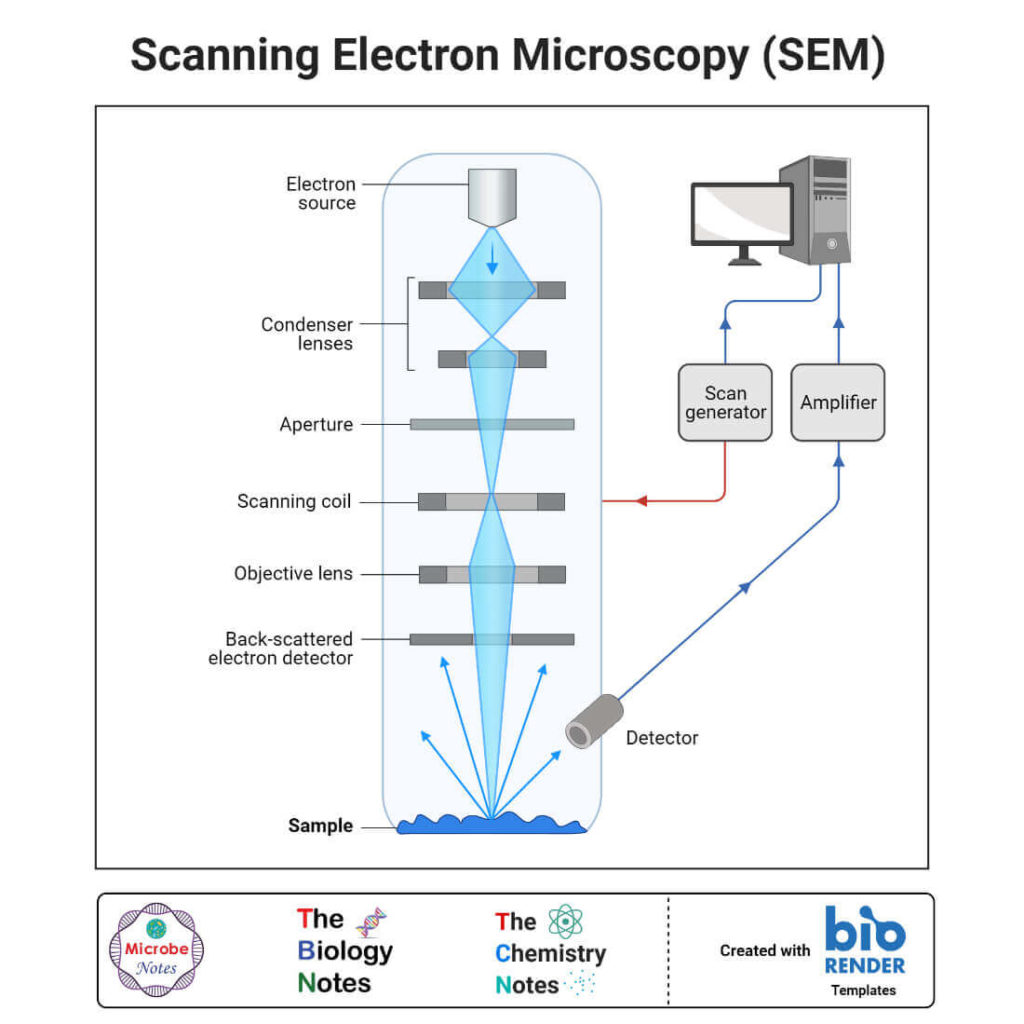
- The image formed at each point during the scanning is combined using a computer to generate an image of a larger region of the sample at a selected magnification.
- The image in such a binocular compound microscope is no different from that obtained with a single monocular eyepiece.
- He sandwiched a very small glass ball lens between the holes in two metal plates riveted together and with an adjustable by screws needles attached to the mouth of the specimen.
- This allows you to view objects and focus on them using your microscope.
The ultrasonic linear motors provide a very wide dynamic speed range from microns/second and below, up to more than 100mm/sec. A linear encoder with 100nm resolution is integrated for each axis – this direct position measurement principle eliminates play and backlash. The U-780 XY stage systems come with many software tools, such as PIMikroMove, PI General Command Set , and drivers for LabVIEW. You will need access to a cell phone that enables you at least to zoom manually in and out. You will need a 1-millimeter optical ball lens to magnify the sample.
How Does A Microscope Achieve Magnification?
Because the objective lens is convex, it focuses and directs light into its center. By contrast, the concave shape of the ocular lens serves to spread out the light as it meets the eye, thereby making the image bigger. The condenser is a lens, often implanted into the stage or located just below it, that condenses the light rays from the light source onto the spot that is being examined on the specimen above.
Step 3: 3d Print The Tube
Generally high vapour solvents are used in order to limit contact time with surfaces and cements. Caution is required when dealing with older microscopes if too much solvent is applied then this may affect the integrity of the mountant e.g. cement or balsam in particular. With modern microscopes this is less of a problem due to the use of polyacrylamates and other more resistant substances.
Modern Photonics Microscope Lens Adapters
In the gold-standard case, it is approximately 180 × 180 μm for 1.25 NA, 100 × oil immersion microscope objective. Current WHO standard requires 100 fields of view to provide the final determination of malaria infection. The standard limit of detection is 30 parasites/μl of blood. The system is almost insensitive to the distance between the glass ball and the phone lens.
For starters, a microscope is an optical tool that can be used to view very small and near objects in great detail. Both instruments were invented hundreds of years ago and have been used to make significant advancements in biology, chemistry, and astronomy. The diameter of the lenses of microscopes and telescopes are substantially different. An optical instrument having a magnifying lens or a combination of lenses for inspecting objects too small to be seen distinctly by the unaided eye.
Microscopes use a convex lens to generate extremely magnified images of very small objects. The lens at the end of the simple microscope produces an inverted and magnified image. Biconvex lenses are a simple lens comprising two convex spherical surfaces, generally with the same radius of curvature. … Being a simple lens, biconvex lenses have a broad range of applications including, but not limited to, focussing and control of laser beams, moderate quality imaging and other optical instruments. The VH lens series features high-performance lenses that bring the best observation and imaging out of microscopes.
Cells are building blocks for all living organisms, but they are too small to be seen with our naked eyes. With the aid of microscopes, we can amplify the power to see these tiny cells. There are also many tiny creatures that we can not see with our naked eyes, such as plankton and bacteria. Illustrated in Figure 8 is a schematic drawing of light waves reflecting and/or passing through a lens element coated with two antireflection layers. The incident wave strikes the first layer at an angle, resulting in part of the light being reflected (R) and part being transmitted through the first layer.
